


Department of Mineralogy, Petrology and Deposit Geology
Laboratory of Scanning Electron Microscopy (SEM)
(room G-330)
The laboratory is equipped with a modern scanning electron microscope JEOL JSM‑IT210 (installed at the end of 2025), which represents a new generation of compact and highly automated SEM systems. The instrument enables fast and precise imaging of samples thanks to a motorized five-axis stage and intelligent features such as Simple SEM for automated acquisition and Zeromag for intuitive navigation between optical and SEM images.
The microscope is capable of high resolution scanning and is equipped with an integrated EDS (Energy Dispersive Spectroscopy) system, which enables elemental analysis and the creation of elemental distribution maps. For more detailed analysis, the system offers topographical measurements, line scanning, automated imaging of the entire sample surface or selected areas, as well as 3D mapping, significantly expanding the possibilities for material characterization. The detectable elemental range in EDS analysis covers almost the entire periodic table, from boron (B) to uranium (U). The accelerating voltage can be set in the range of 0.3 to 30 kV.
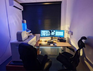
The JSM‑IT210 also supports measurements in low-vacuum (LV) mode, which allows imaging and/or analysis of non-conductive, biological, geological, and otherwise untreated samples without the need for conductive coating. The EDS system is also compatible with low-vacuum mode, enabling the acquisition of high-quality elemental maps even for non-conductive materials. It also offers stable operation with reduced surface charging, which is particularly important for the analysis of polymers, glasses, minerals, and biological tissues.
 |  |
|---|
Within the laboratory, we also offer cathodoluminescence (CL) analysis, which complements the morphological and chemical information obtained from electron microscopy. This technique enables the study of optical properties of materials, the identification of crystal lattice defects, and the zoning of minerals that may not be visible in SEM/BSE imaging. CL analysis is enabled by a combined specialized SEM/CL detector.
The instrument enables the following for the study of minerals, geological and biological samples, and other materials:
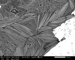
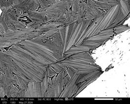
- backscattered electron (BSE) imaging displaying differences in the chemical composition of geomaterials
- surface imaging in secondary electron (SE) mode
- chemical composition analysis using EDS (Energy Dispersive Spectroscopy) from boron (B) to uranium (U)
- elemental distribution maps in EDS mode
- topography measurements, line scans, 3D mapping, and area-based compositional analyses of the studied material
- cathodoluminescence (CL) imaging of cathodoluminescent minerals and materials
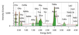
The instrument is controlled by the InTouchScope™ software platform with the additional Smile View software package for processing photographs, montages, topographic data, and analyzed EDS spectra. The laboratory includes a QUORUM Q150R ESplus carbon sputtering machine for preparing samples in an electron microscope.
Costs for internal accounting, for external customers and commercial institutions (PDF)