


Chaetonotus (Hystricochaetonotus) avarus
Reference
Rataj Križanová F. & Vďačný P. (2022) A huge undescribed diversity of the subgenus Hystricochaetonotus (Gastrotricha, Chaetonotidae, Chaetonotus) in Central Europe. European Journal of Taxonomy 840: 1-93. https://doi.org/10.5852/ejt.2022.840.1941
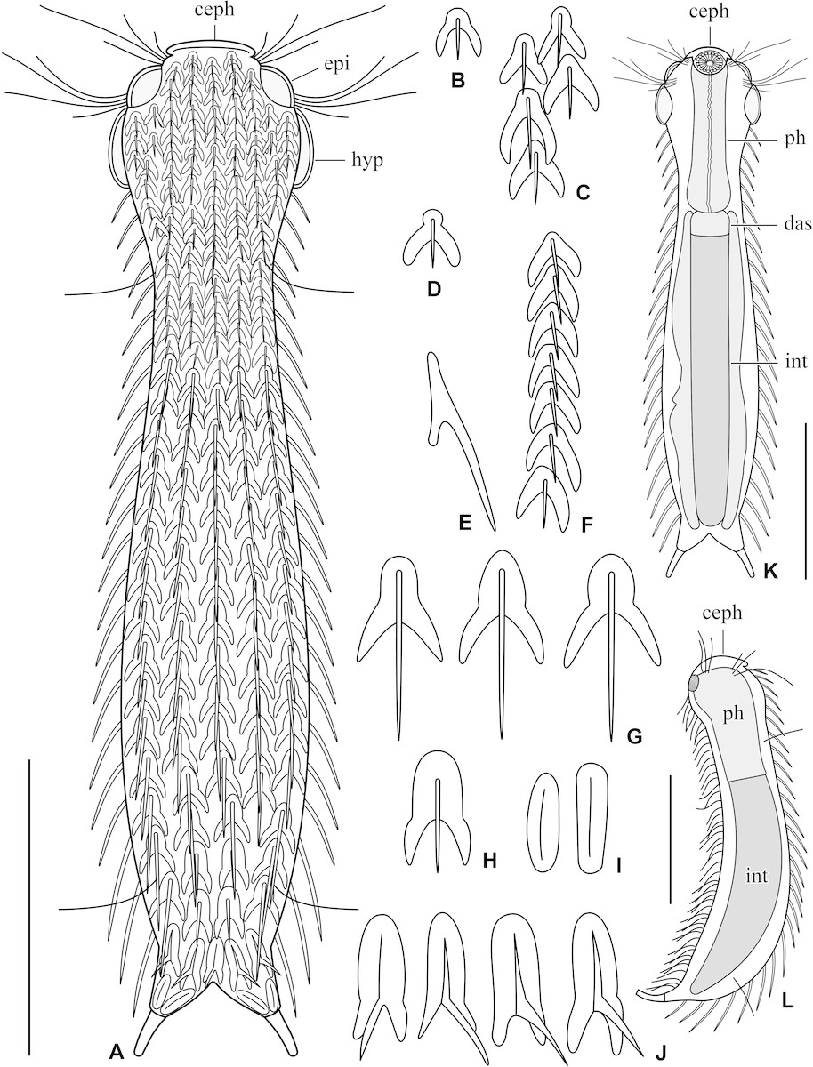
Chaetonotus (Hystricochaetonotus) avarus, schematic drawings. (A) Dorsal overview showing the scale and spine pattern. (B–C) Head scales. (D) Neck dorsolateral scale. (E) Lateral view of a trunk spined scale. (F) Neck central scales. (G) Trunk scales. (H) Posterior trunk scales. (I) Dorsal furca branches scales. (J) Dorsal furca base scales. (K) Internal morphology. (L) Lateral overview. Scale bars: 30 μm.
© 2022 Rataj Križanová F. & Vďačný P. This picture is licensed under a CC BY-NC-ND 4.0 license.
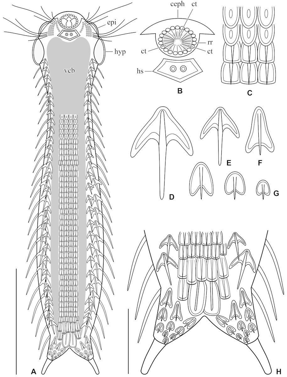
Chaetonotus (Hystricochaetonotus) avarus, schematic drawings. (A) Ventral overview showing the scale and ciliary pattern. (B) Mouth region. (C) Detail showing the transition region of two scales types in the interciliary field. (D–E) Ventrolateral three-lobed scales. (F) Ventral furcal three-lobed scales. (G) Furcal cordiform scales. (H) Detail of the terminal ventral region. Scale bars: 10 µm (H) and 30 µm (A).
© 2022 Rataj Križanová F. & Vďačný P. This picture is licensed under a CC BY-NC-ND 4.0 license.
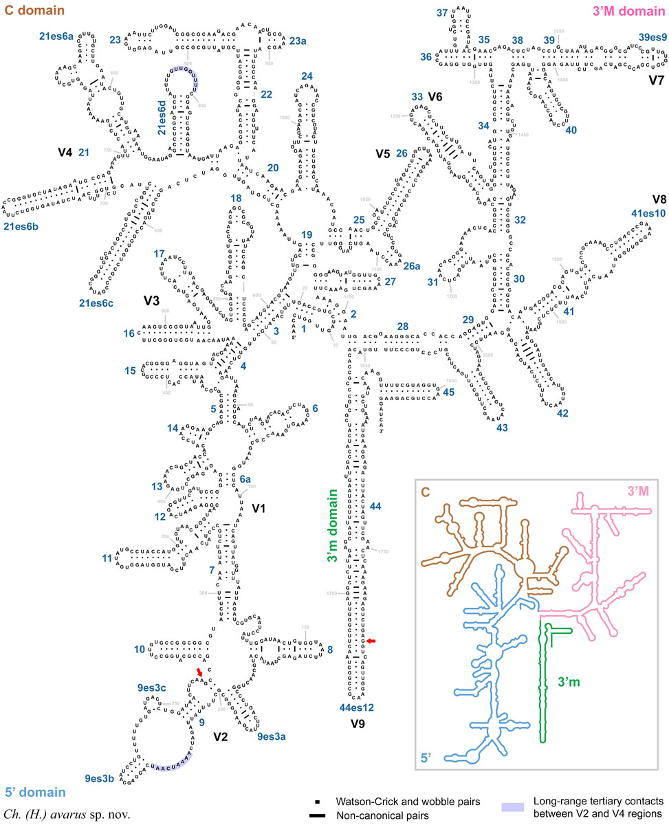
Secondary structure of the 18S rRNA molecule of Chaetonotus (Hystricochaetonotus) avarus. Diagnostic molecular autapomorphies are marked by red arrows. The reference 18S secondary structure map of Saccharomyces cerevisiae (inset) is from apollo.chemistry.gatech.edu/RibosomeGallery.
© 2022 Rataj Križanová F. & Vďačný P. This picture is licensed under a CC BY-NC-ND 4.0 license.
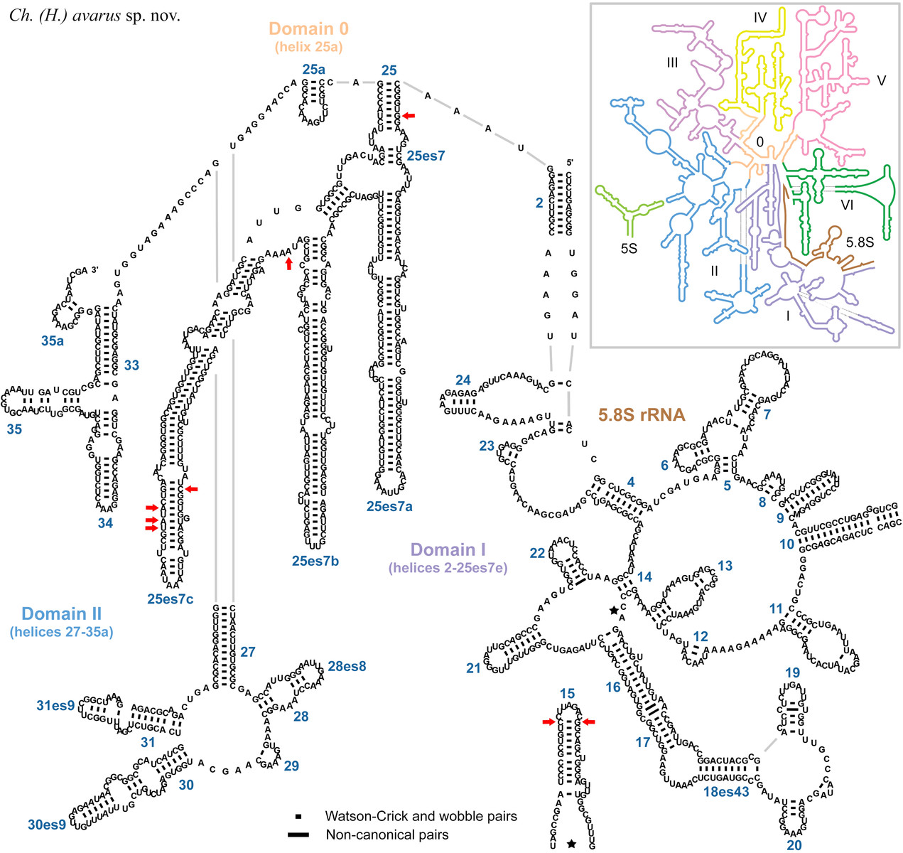
Secondary structure of the first two domains of the 28S rRNA molecule of Chaetonotus (Hystricochaetonotus) avarus. Diagnostic molecular autapomorphies are marked by red arrows. The reference 28S secondary structure map of Saccharomyces cerevisiae (inset) is from apollo.chemistry.gatech.edu/RibosomeGallery.
© 2022 Rataj Križanová F. & Vďačný P. This picture is licensed under a CC BY-NC-ND 4.0 license.
Morphological diagnosis
Body elongated and about 130 μm long. Head slightly wider than neck, separated from trunk by an inconspicuous neck constriction. Cephalion, epipleurae, and hypopleurae clearly demarcated. Trunk comparatively narrow, not broader than head, widest at ca U62, narrowest at ca U87. Mouth ventral, three cuticular teeth. Hypostomium bears a pentagonal cuticular structure with two protuberances. Pharynx without dilatations. Intestine straight, with a marked anterior section. Scales slightly overlapping, distributed in 14 columns, 30–32 scales per column. Dorsal surface covered with: (i) head scales small, with a subtle, well-delimited anterior lobe and comparatively long and diverging posterior lobes; (ii) neck scales boomerang-like, with a broadly rounded anterior lobe, elongated posterior lobes; (iii) dorsolateral neck scales with a well-delimited, broadly rounded anterior lobe and distinct transition between anterior and posterior lobes; (iv) main trunk scales with a rounded anterior end, posterior lobes comparatively long, narrowly rounded distally, and diverging; (v) posterior trunk scales with a conspicuously prolonged, tongue-shaped anterior lobe and relatively short posterior lobes tapering distally; and (vi) furca base scales with a narrower and more elongated anterior lobe and short posterior lobes very narrowly rounded or almost acute distally. Spines slightly increasing in length from head to posterior trunk region, gradually tapering to become hair-like terminally, without denticles. Ventrolateral spines shorter than dorsal ones. Ventral side carries two broad, anteriorly connected (U9–U27) ciliary bands. Interciliary field covered by (i) minute, tongue-like, keeled scales; (ii) small, roughly rectangular, keeled scales; (iii) oblong scales with a short spine; and (iv) a pair of big, elongated oval, keeled scales. Furca branches slightly longer than adhesive tubes, lateral margins more or less straight, furcal indentation deeply V-shaped, adhesive tubes short. Dorsal side of furca branches covered with elongated oval, keeled scales and ventral side of furca branches covered with (i) two pairs of three-lobed, spined scales, having an elongated, tongue-shaped anterior lobe and comparatively short, narrowly rounded to acute posterior lobes and (ii) cordiform, short-spined scales.
Molecular diagnosis
18S rRNA gene: 268 A, 1739 G.
28S rRNA gene: 128 C, 136 G, 447 G, 691 A, 737 C, 764 T, 765 A, 767 A.
Cytochrome c oxidase subunit I (codon ordinal numbers are followed by the corresponding span of nucleotide positions in parentheses): 84 (250‒252) CTA, 91 (271‒273) CTA, 99 (295‒297) CTA, 102 (304‒306) TCG, 120 (358‒360) TCC, 154 (460‒462) ACT, 192 (574‒576) GGA, 213 (637‒639) GGG.
Type material
A DNA sample of the holotype specimen VP 32 has been deposited in the Natural History Museum, Vajanského nábrežie 2, 810 06 Bratislava, Slovakia (ID Collection Code 01427566).
Type locality
Vajspeterský potok creek, Rača, Bratislava, Podunajská rovina plain, Slovakia, 48°12′12.8″ N, 17°07′46.9″ E.